

Journal of Clinical Pediatrics ›› 2026, Vol. 44 ›› Issue (5): 418-423.doi: 10.12372/jcp.2026.25e1605
• Original Article • Previous Articles Next Articles
DONG Ying, CAO Tingting, WU Jianping, ZHANG li, GE Hongsong( )
Received:2025-12-16
Accepted:2026-03-17
Published:2026-05-15
Online:2026-05-08
CLC Number:
DONG Ying, CAO Tingting, WU Jianping, ZHANG li, GE Hongsong. Dermoscopic characteristics analysis of acral volar melanocytic nevi in children and adolescents[J].Journal of Clinical Pediatrics, 2026, 44(5): 418-423.
Figure 1
Four variants of the parallel furrow pattern"
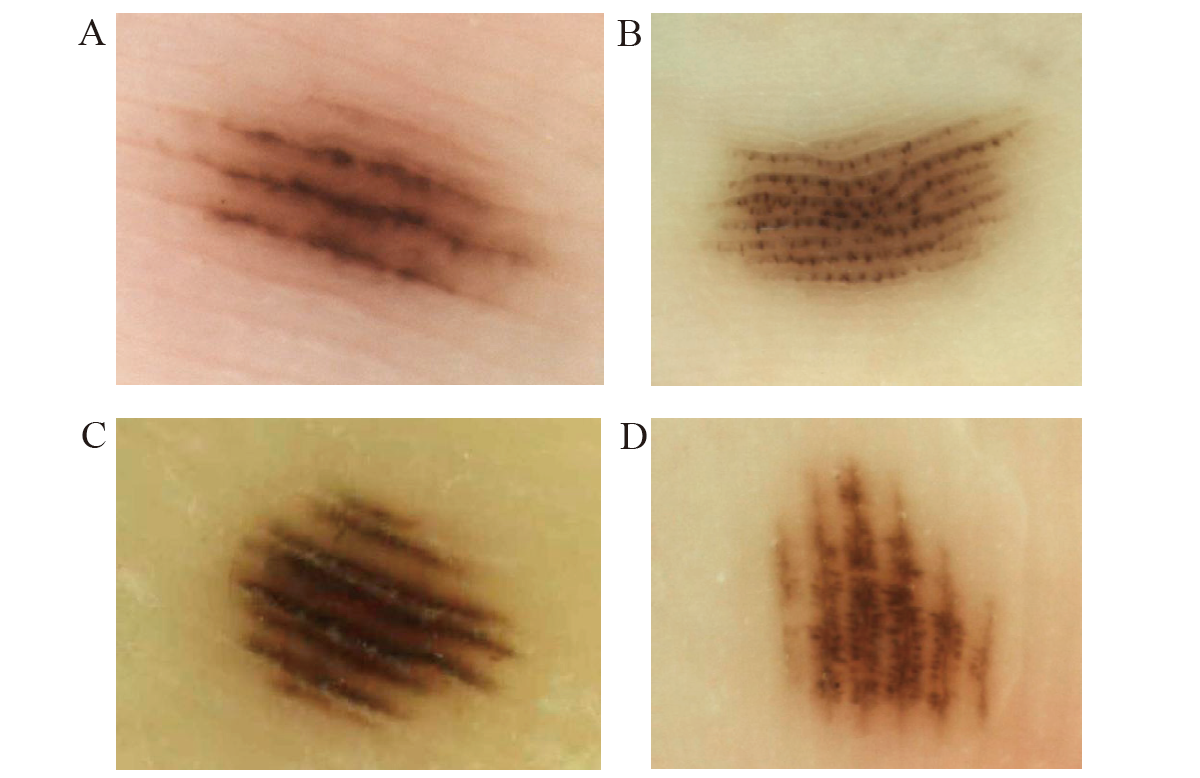
Figure 2
Various dermoscopic patterns of acral volar melanocytic nevus"
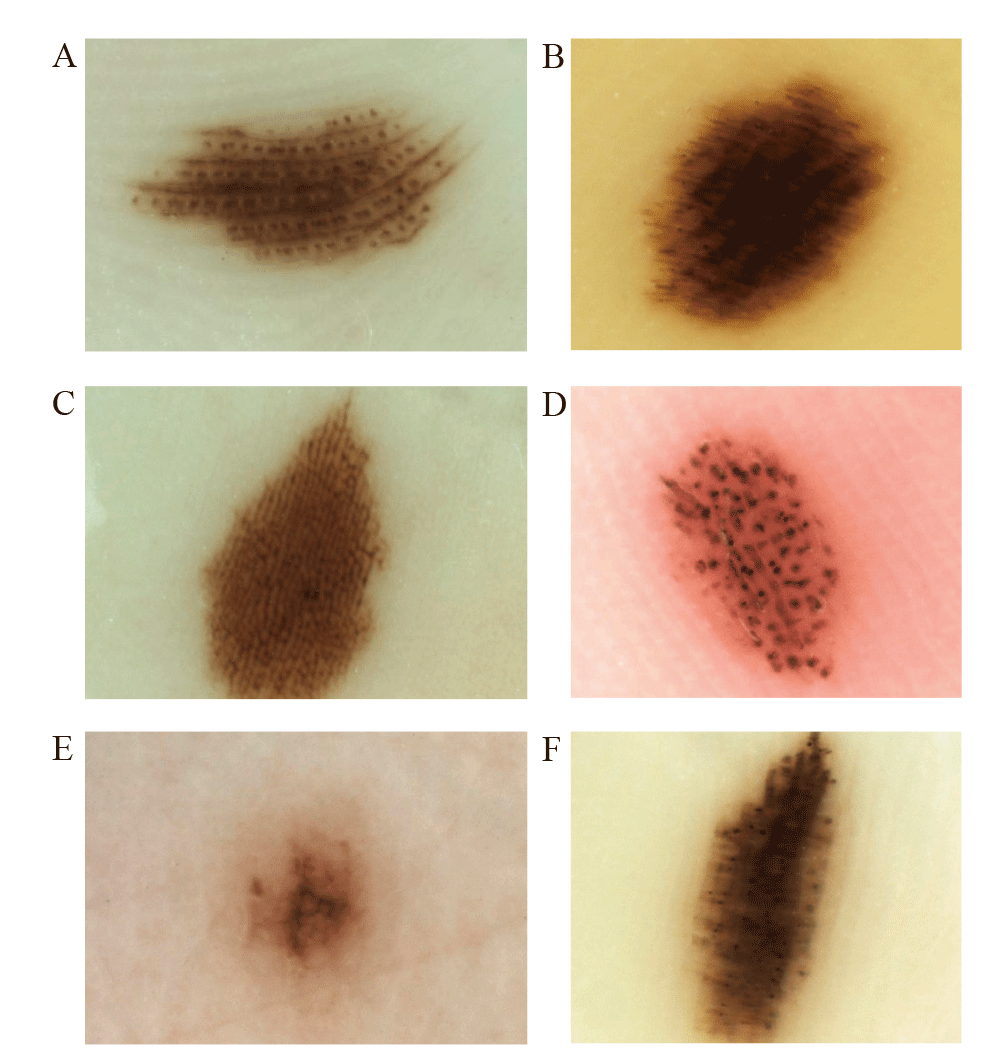

Table 1
Distribution of dermoscopic patterns of 138 acral volar melanocytic nevi in different locations [n(%)]"
| 部 位 | 皮肤镜模式 | 总计 | ||||||||
|---|---|---|---|---|---|---|---|---|---|---|
| 皮沟平行模式 | 网格样模式 | 纤维状模式 | 网状模式 | 球状模式 | 均质模式 | 豌豆荚模式 | 复合模式 | 不典型模式 | ||
| 跖骨区 | 10(32.3) | 2(6.5) | 10(32.3) | 0(0) | 1(3.2) | 0(0) | 5(16.1) | 2(6.5) | 1(3.2) | 31(22.5) |
| 足跟部 | 7(24.1) | 0(0) | 12(41.4) | 0(0) | 0(0) | 0(0) | 6(20.7) | 2(6.9) | 2(6.9) | 29(21.0) |
| 足中部外侧区 | 2(18.2) | 0(0) | 2(18.2) | 0(0) | 0(0) | 0(0) | 6(54.5) | 1(9.1) | 0(0) | 11(8.0) |
| 足弓部 | 4(23.5) | 6(35.3) | 2(11.8) | 0(0) | 1(5.9) | 0(0) | 2(11.8) | 0(0) | 2(11.8) | 17(12.3) |
| 足趾跖侧 | 6(60.0) | 0(0) | 2(20.0) | 0(0) | 0(0) | 0(0) | 1(10.0) | 0(0) | 1(10.0) | 10(7.2) |
| 足底侧缘 | 0(0) | 0(0) | 0(0) | 0(0) | 0(0) | 0(0) | 1(100.0) | 0(0) | 0(0) | 1(0.7) |
| 手指掌侧 | 7(33.3) | 1(4.8) | 0(0) | 0(0) | 4(19.0) | 0(0) | 9(42.9) | 0(0) | 0(0) | 21(15.2) |
| 近端掌部 | 2(50.0) | 1(25.0) | 0(0) | 0(0) | 1(25.0) | 0(0) | 0(0) | 0(0) | 0(0) | 4(2.9) |
| 掌中部 | 0(0) | 0(0) | 0(0) | 0(0) | 2(40.0) | 0(0) | 0(0) | 1(20.0) | 2(40.0) | 5(3.6) |
| 远端掌部 | 0(0) | 0(0) | 0(0) | 0(0) | 0(0) | 0(0) | 0(0) | 0(0) | 1(100.0) | 1(0.7) |
| 大鱼际 | 3(60.0) | 0(0) | 0(0) | 0(0) | 0(0) | 0(0) | 1(20.0) | 1(20.0) | 0(0) | 5(3.6) |
| 小鱼际 | 2(66.7) | 0(0) | 0(0) | 0(0) | 0(0) | 0(0) | 1(33.3) | 0(0) | 0(0) | 3(2.2) |
| 手掌侧缘 | 0(0) | 0(0) | 0(0) | 0(0) | 0(0) | 0(0) | 0(0) | 0(0) | 0(0) | 0(0) |
| 总计 | 43(31.2) | 10(7.2) | 28(20.3) | 0(0) | 9(6.5) | 0(0) | 32(23.2) | 7(5.1) | 9(6.5) | 138 |
Table 2
The correlation between dermoscopic patterns and anatomic localization of acral volar melanocytic nevi [n(%)]"
| 皮肤镜模式 | 例数 | 足弓 | 足底承重部位 | 足底其他部位 | 手指掌侧 | 手掌其他部位 | χ2值 | P |
|---|---|---|---|---|---|---|---|---|
| 皮沟平行模式 | 43 | 4(9.3) | 19(44.2) | 6(14.0) | 7(16.3) | 7(16.3) | 50.78 | <0.001 |
| 纤维状模式 | 28 | 2(7.1) | 24(85.7) | 2(7.1) | 0(0) | 0(0) | ||
| 网格样模式 | 10 | 6(60.0)1)2) | 2(20.0)2) | 0(0) | 1(10.0) | 1(10.0) | ||
| 豌豆荚模式 | 32 | 2(6.3)3) | 17(53.1) | 2(6.3) | 9(28.1)2) | 2(6.3) | ||
| 其他模式 | 25 | 3(12.0) | 9(36.0) | 1(4.0) | 4(16.0) | 8(32.0)2) |
| [1] |
Saida T, Oguchi S, Ishihara Y. In vivo observation of magnified features of pigmented lesions on volar skin using video macroscope. Usefulness of epiluminescence techniques in clinical diagnosis[J]. Arch Dermatol, 1995, 131(3): 298-304.
pmid: 7887659 |
| [2] |
González-Ramírez RA, Guerra-Segovia C, Garza-Rodríguez V, et al. Dermoscopic features of acral melanocytic nevi in a case series from Mexico[J]. An Bras Dermatol, 2018, 93(5): 665-670.
doi: S0365-05962018000500665 pmid: 30156615 |
| [3] |
Savas Erdogan S, Falay Gur T, Turgut Erdemir AV, et al. Dermoscopic characteristics of acral melanocytic nevi in children and adolescents[J]. Pediatr Dermatol, 2020, 37(4): 597-603.
doi: 10.1111/pde.14136 pmid: 32291825 |
| [4] | Marghoob AA, Malvehy J, Braun RP. 皮肤镜图谱[M]. 第二版. 徐峰, 等译. 上海: 复旦大学出版社, 2018: 268-275. |
| [5] |
Park S, Yun SJ. Acral melanocytic neoplasms: a comprehensive review of acral nevus and acral melanoma in Asian perspective[J]. Dermatopathology (Basel), 2022, 9(3): 292-303.
doi: 10.3390/dermatopathology9030035 pmid: 35997352 |
| [6] |
Suzaki R, Ishizaki S, Iyatomi H, et al. Age-related prevalence of dermatoscopic patterns of acral melanocytic nevi[J]. Dermatol Pract Concept, 2014, 4(1): 53-57.
doi: 10.5826/dpc.0401a08 pmid: 24520515 |
| [7] |
Saida T, Koga H. Dermoscopic patterns of acral melanocytic nevi: their variations, changes, and significance[J]. Arch Dermatol, 2007, 143 (11): 1423-1426.
doi: 10.1001/archderm.143.11.1423 pmid: 18025367 |
| [8] |
Wawrzynkiewicz M, Pastuszczak M, Chlebicki M, et al. Acral melanocytic nevi in the Polish population: prevalence, clinical characteristics and dermoscopic patterns[J]. Postep Derm Alergol, 2019, 36 (5): 581-588.
doi: 10.5114/ada.2018.77500 pmid: 31839775 |
| [9] |
Roh D, Shin K, Kim WI, et al. Comparison of dermoscopic features between congenital and acquired acral melanocytic nevi in Korean patients[J]. J Eur Acad Dermatol, 2020, 34(5): 1004-1009.
doi: 10.1111/jdv.v34.5 |
| [10] | Karaca Ural Z, Baykal Selcuk L, Aksu Arica D, et al. Prevalence and dermoscopic patterns of acral melanocytic nevi in Turkey[J]. Acta Dermatovener Cr, 2021, 29(2): 94-101. |
| [11] |
Minagawa A, Koga H, Saida T. Dermoscopic characteristics of congenital melanocytic nevi affecting acral volar skin[J]. Arch Dermatol, 2011, 147(7): 809-813.
doi: 10.1001/archdermatol.2011.150 pmid: 21768479 |
| [12] |
Minagawa A, Koga H, Uhara H, et al. Age-related prevalence of dermoscopic patterns in acquired melanocytic nevus on acral volar skin[J]. JAMA Dermatol, 2013, 149(8): 989-990.
doi: 10.1001/jamadermatol.2013.4452 pmid: 23804247 |
| [13] |
Emiroglu N, Cengiz FP, Onsun N. Age and anatomical location-related dermoscopic patterns of 210 acral melanocytic nevi in a Turkish population[J]. J Cutan Med Surg, 2017, 21(5): 388-394.
doi: 10.1177/1203475417712496 pmid: 28565919 |
| [14] |
Miyazaki A, Saida T, Koga H, et al. Anatomical and histopathological correlates of the dermoscopic patterns seen in melanocytic nevi on the sole: a retrospective study[J]. J Am Acad Dermatol, 2005, 53(2): 230-236.
doi: 10.1016/j.jaad.2005.04.045 pmid: 16021115 |
| [15] |
Maumi Y, Kimoto M, Kobayashi K, et al. Oblique view dermoscopy changes regular fibrillar pattern into parallel furrow pattern[J]. Dermatology, 2009, 218(4): 385-386.
doi: 10.1159/000202986 |
| [16] | Tognetti L, Cartocci A, Moscarella E, et al. Pattern analysis of benign and malignant atypical melanocytic skin lesions of palms and soles: variations of dermoscopic features according to anatomic site and personal experience[J]. Life (Basel), 2024, 14(6): 659. |
| [17] | Barquet V, Dufrechou L, Nicoletti S, et al. Dermoscopic patterns of 158 acral melanocytic nevi in a Latin American population[J]. Actas Dermo-Sifilogr, 2013, 104(7): 586-592. |
| [18] |
Papageorgiou C, Kyrgidis A, Ilut PA, et al. Acral melanocytic nevi in a high-risk population: prevalence, clinical characteristics and dermatoscopic patterns[J]. Dermatology, 2023, 239(5): 753-759.
doi: 10.1159/000531055 |
| [19] |
Ingrassia JP, Stein JA, Levine A, et al. Diagnosis and management of acral pigmented lesions[J]. Dermatol Surg, 2023, 49(10): 926-931.
doi: 10.1097/DSS.0000000000003891 pmid: 37556446 |
| [20] | Saida T, Koga H, Uhara H. Dermoscopy for acral melanocytic lesions: revision of the 3-step algorithm and refined definition of the regular and irregular fibrillar pattern[J]. Dermatol Pract Concept, 2022, 12(3): e2022123. |
| [1] | LIN Jinfeng, CHEN Yi, DENG Yan, FENG Ai, HUANG Juan, CHEN Lizhu, XIE Jingqi, NIE Xiaojing. A comparative study on the detection of anti-Nephrin antibodies in children's nephrotic syndrome by chemiluminescence method and cell-based assay [J]. Journal of Clinical Pediatrics, 2026, 44(8): 682-689. |
| [2] | ZHENG Fangyuan, WANG Miao, DING Mingming, LU Aidong, JIA Yueping, ZENG Huimin, ZHANG Leping. Next-generation sequencing identified gene variants and their prognostic impact in pediatric Philadelphia chromosome positive acute lymphoblastic leukemia [J]. Journal of Clinical Pediatrics, 2026, 44(8): 690-697. |
| [3] | ZHANG Xiao, FANG Hongjun, KUANG Xiaojun, WANG Lijuan, WU Zhao, JIANG Sha, LI Pei, WU Liwen. Analysis of the clinical and electrophysiological characteristics of atonic seizures in children [J]. Journal of Clinical Pediatrics, 2026, 44(8): 698-704. |
| [4] | WANG Mengqian, YANG Hui. Case series analysis of 13 intestinal Behcet’s syndrome in children [J]. Journal of Clinical Pediatrics, 2026, 44(8): 705-711. |
| [5] | WU Xirong, GAO Qi, GUO Siyuan, YIN Qingqin, QIN Qiang, YIN Ju, JIAO Anxia, CHEN Chenghao, WU Di, WANG Bei, DUAN Xiaomin, PENG Yun, XU Baoping, GAO Liwei. Clinical characteristics and prognosis of pulmonary mucormycosis in children with diabetes mellitus [J]. Journal of Clinical Pediatrics, 2026, 44(8): 712-720. |
| [6] | HAN Yanan, GE Lanlan, CUI Jieyuan. A case report of branchio-oto-renal syndrome caused by EYA1 gene variant with a 6-year follow-up [J]. Journal of Clinical Pediatrics, 2026, 44(8): 721-726. |
| [7] | SHI Jiayi, GAO Chunlin, JIA Lili, LU Yunyun, SUN Tao, ZHU Xiaodong, XIA Zhengkun, ZHANG Pei. Phenotypic characteristics and prognostic analysis of a pedigree with childhood isolated nephrotic syndrome caused by compound heterozygous mutations in the COQ2 gene [J]. Journal of Clinical Pediatrics, 2026, 44(8): 727-732. |
| [8] | Rachel Rosen, Osvaldo Borrelli, Christophe Faure, Katja Karrento, Usha Krishnan, Samuel Nurko, Nathalie Rommel, Alan Silverman, Michiel van Wijk, Marc Benninga (Author), Chinese Medical Association Parenteral and Enteral Nutrition Branch Pediatrics Group, The Subspecialty Group of Gastroenterology, The Society of Pediatric, Chinese Medical Association, Gastroenterology Group, Pediatrician Branch, Chinese Medical Doctor Association (Translator). Core interpretation and explanation of Rome Ⅴ diagnostic criteria: upper gastrointestinal disorders of gut-brain interaction in children and adolescents [J]. Journal of Clinical Pediatrics, 2026, 44(7): 595-608. |
| [9] | Carlo Di Lorenzo, Miguel Saps, Bruno P. Chumpitazi, Shaman Rajindrajith, Annamaria Staiano, Nikhil Thapar, Miranda van Tilburg, Carlos Velasco-Benítez, Arine Vlieger (Author), Chinese Medical Association Parenteral and Enteral Nutrition Branch Pediatrics Group, The Subspecialty Group of Gastroenterology, The Society of Pediatric, Chinese Medical Association, Gastroenterology Group, Pediatrician Branch, Chinese Medical Doctor Association (Translator). Core interpretation and explanation of Rome Ⅴ diagnostic criteria: lower and biliary disorders of gut-brain interaction in children and adolescents [J]. Journal of Clinical Pediatrics, 2026, 44(7): 609-621. |
| [10] | LI Xufeng, TANG Haoxun, LI Xingjun, ZHANG Lize, GAO Chenqing, ZHAO Shunying, LIU Jianhua. Clinical characteristics, chest CT imaging phenotypes, and their correlation with prognosis in 76 children with Chlamydia pneumoniae pneumonia [J]. Journal of Clinical Pediatrics, 2026, 44(7): 622-627. |
| [11] | WANG Zhen, SHAO Jingbo, ZHANG Na, XIA Min, ZHU Jiashi, DU Chengkan, LI Hong. Factors influencing long-term recurrence in pediatric acute lymphoblastic leukemia patients with low-risk disease at initial diagnosis: a five-year follow-up analysis of 254 cases from a single center [J]. Journal of Clinical Pediatrics, 2026, 44(7): 628-635. |
| [12] | GUO Xuemei, XUE Yao, WANG Yongren, RONG Liucheng, FANG Yongjun, LIN Rufeng. Clinical features and prognosis of childhood acute myeloid leukemia with CBFA2T3::GLIS2 fusion [J]. Journal of Clinical Pediatrics, 2026, 44(7): 636-643. |
| [13] | YANG Lijun, FU Dongxia, CUI Yan, YANG Junmei, ZHANG Liming. Clinical manifestation and genetic analysis of CHARGE syndrome caused by CHD7 gene variation [J]. Journal of Clinical Pediatrics, 2026, 44(7): 644-650. |
| [14] | HU Yan, YANG Fan, LIANG Aimin, MAO Meng. Identification and management of faltering growth in children: insights from the 13th International Conference on Nutrition and Growth [J]. Journal of Clinical Pediatrics, 2026, 44(6): 485-488. |
| [15] | LIU Ziyao, WANG Congying, WANG Hongmao, ZHANG Mingming, XU Yingjie, LAI Jianming, NIU Wenquan, LI Xiaohui. Early differentiation of Kawasaki disease with joint symptoms and systemic juvenile idiopathic arthritis with Kawasaki disease-like symptoms: a single-center retrospective study [J]. Journal of Clinical Pediatrics, 2026, 44(6): 489-498. |
|
||
